
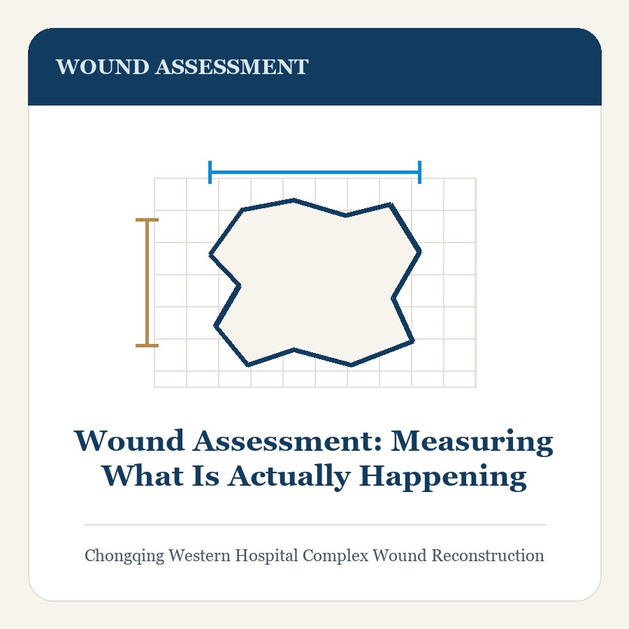
In short. How clinicians assess a wound: measurement, tissue type, infection, perfusion, and sensation, and how a structured record shows whether treatment is working.
Key points
- A wound can look cleaner while it is getting larger, and can look alarming while it is shrinking steadily.
- That is why area and depth come before colour in a proper assessment, and why serial photographs taken the same way each time — same distance, same light, same scale marker — are more useful than a single impressive image.
Good wound care begins with a record. Without one, every visit becomes a fresh opinion, and slow deterioration can hide behind a dressing. A structured assessment takes only minutes, but it is what turns wound care from guesswork into a plan that can be judged on evidence.
Measuring the wound
Length, width, and depth are measured in centimetres at each visit, using the same clock position and the same patient position every time. Area is tracked over weeks, and depth is probed gently to detect undermining and tunnelling, where the wound extends beneath intact skin. Two numbers matter more than appearance: whether the area is shrinking, and whether depth is decreasing.
Tissue type in the wound bed
What sits at the base of the wound predicts how it will behave. Pink granulation tissue means healing is under way. Yellow slough and black eschar are dead tissue that physically blocks repair and hides infection underneath. Undermined edges that roll inward, or edges that feel hard, suggest the wound has stalled and needs more than a dressing change.
Measure first, then look
A wound can look cleaner while it is getting larger, and can look alarming while it is shrinking steadily. That is why area and depth come before colour in a proper assessment, and why serial photographs taken the same way each time — same distance, same light, same scale marker — are more useful than a single impressive image.
Infection
Classic signs are spreading redness, warmth, pain, swelling, and pus. Chronic wounds often show subtler signs: new pain, a friable surface that bleeds easily, delayed healing despite good care, or an unpleasant odour. A surface swab mostly grows colonising organisms and misleads; deep tissue sampling or culture of drained pus is needed when infection is genuinely suspected.
Perfusion and sensation
Pulses, skin colour, temperature, hair growth, and capillary refill give a first impression of blood supply. Ankle–brachial index measurement and, where needed, toe pressures or duplex imaging classify the circulation properly, because that result decides whether compression is safe or dangerous. Sensation testing with a fine monofilament detects the neuropathy that lets diabetic foot wounds go unnoticed until they are deep.
Documenting progress
The assessment is recorded and compared over time. If a wound has not reduced in area after four weeks of correct treatment, the diagnosis is revisited rather than the dressing changed again. Failure to improve is information: it usually means that pressure, perfusion, infection, or diabetes control has not yet been addressed.
Seek urgent review if
- The wound enlarges or deepens, or begins to expose bone, tendon, or joint.
- Redness spreads, or fever, foul odour, or pus develops.
- Pain becomes severe or new, or a limb turns cold, pale, or blue.
Frequently asked questions
Why does a wound need measuring at every visit?
Area and depth tracked over weeks show whether treatment is working, and they matter more than appearance. A wound can look cleaner while enlarging, or look alarming while shrinking. Measurements taken at the same clock position and patient position make that visible.
What do the different tissue types in a wound bed mean?
Pink granulation tissue means healing is under way. Yellow slough and black eschar are dead tissue that blocks repair and can hide infection beneath them. Rolled or hard wound edges suggest the wound has stalled and needs more than a dressing change.
Does a swab from the wound surface show the infection?
Rarely in a useful way. A surface swab mostly grows colonising organisms and biofilm, which can prompt unnecessary antibiotics. When infection is genuinely suspected, deep tissue taken during debridement, or pus from a collection, is the useful sample.
Risks & Limitations
- Chronic wounds heal over months, not weeks. A wound that closes in three to six months of consistent care is a good result.
- Recurrence is common. Both diabetic foot wounds and venous leg ulcers frequently return, which is why protective footwear and compression continue after healing.
- Partial or complete failure of a skin graft or flap, which may require further surgery.
- Infection and biofilm, which delay healing by weeks and can spread deeply or into bone.
- Amputation risk in diabetes and vascular disease, which is reduced but never abolished by treatment.
- Prolonged dressings and repeated clinic visits. Offloading, compression, and follow-up are demanding and take months.
- Donor-site scarring and discomfort where a graft or flap was taken.
- Reduced or absent sensation in grafted and flap tissue, and around a healed diabetic foot wound.
When to Seek Care
- A wound that is enlarging or deepening, or that begins to expose bone, tendon, or joint.
- Black, grey, or dead tissue appearing in or around the wound.
- Spreading redness, warmth, or swelling around the wound.
- Fever, shivering, or feeling generally very unwell.
- Foul odour or pus from the wound.
- New or severe pain, including pain in the leg or foot at rest or at night.
- A toe or foot that turns black, cold, or blue.
- Loss of sensation in a foot or toe, or a new wound or blister you cannot feel.
References & Further Reading
- World Health Organization (WHO) — chronic wound and diabetes care resources
- NICE (National Institute for Health and Care Excellence) — guidance on wound care, diabetic foot problems, and pressure ulcers
- Wounds International — international consensus documents on wound infection, biofilm, and compression therapy
- American Society of Plastic Surgeons (ASPS) — plastic surgery guidance on wound closure
- Cochrane Library — systematic reviews in wound care and wound closure
This page is patient education, not medical advice, and it does not cite individual studies. Discuss your own case with a qualified clinician. To arrange an assessment at Chongqing West District Hospital, see Contact & Consultation.